MRI-DTI
Professor Emeritus UCI Medical School Joseph C. Wu M.D.
A New Paradigm
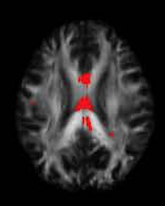
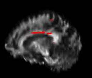
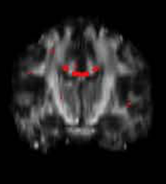
MRI-DTI is a newer MRI sequence that is much more sensitive at detecting signs of axonal shearing due to brain trauma than conventional CT or MRI imaging.
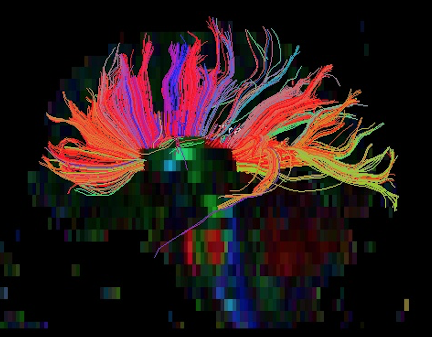
Tractography
Visualizations of the corpus callosum or other fiber tracts show abnormalities due to tearing of the axons. Z-Map analysis shows abnormalities in fractional anisotropy due to damage from injury.
Imaging Site Options
HOAG Hospital Newport Beach
HOAG has 3 Tesla MRI-DTI capabilities and is located nearby.
Other MRI Facilities
We also accept and analyze statistically MRI-DTI scans from most other facilities. Please contact us for details.
Insights
TBI
TBI's can be detected with MRI-DTI by measuring a property called fractional anisotropy (FA) and comparing the FA values of different voxels in the brain to normal controls.
Hypoxia
Hypoxia from Carbon Monoxide poisoning or near-drowning events can cause significant aberrations in FA on MRI-DTI.
Schizophrenia
Schizophrenia is associated with significant reduction in FA values in regions such as the corpus callosum.
Gallery
Quantitative image analysis provides objective statistical measurements of brain abnormalities in brain injury cases, which are often overlooked by conventional radiologists. Feel free to reach out.
Contact us